
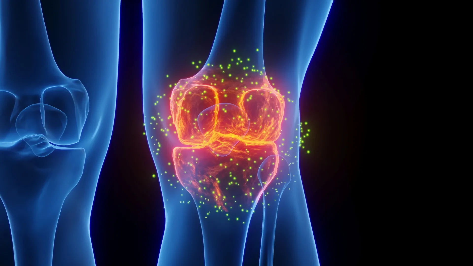
For millions of people living with osteoarthritis, the daily reality is defined by a slow, creeping decline in mobility accompanied by persistent pain and joint stiffness. Standard therapeutic options—ranging from over-the-counter pain medications and non-steroidal anti-inflammatory drugs to localized steroid injections—are fundamentally limited. While these conventional treatments may offer temporary relief from acute symptoms, they do little to alter the underlying trajectory of the disease, leaving the structural degradation of the joint unchecked as the condition steadily advances.
Now, a breakthrough study conducted by researchers at Yale University points toward an entirely new therapeutic paradigm. Published in the journal Bioactive Materials, the research demonstrates that the medication lacosamide—traditionally prescribed to manage epilepsy—can fulfill a dual purpose in the context of osteoarthritis. According to the findings, the drug is capable not only of dampening debilitating joint pain but also of actively reversing cartilage damage. Crucially, these regenerative and analgesic effects were significantly amplified when the medication was administered directly into the joint cavity via a newly engineered, temperature-responsive hydrogel.
Why Osteoarthritis Damages Cartilage
Osteoarthritis is frequently described in colloquial terms as a simple condition caused by wear and tear, but medical specialists emphasize that this characterization overlooks the complex biological mechanisms driving the disease.
Within a structurally sound, healthy joint, specialized cells known as chondrocytes assume the vital responsibility of maintaining the integrity of the cartilage. They achieve this by constantly regulating a delicate balance between the creation of new extracellular matrix tissue and the enzymatic removal of aged or damaged material. Osteoarthritis fundamentally disrupts this equilibrium. Under pathological conditions, the cartilage begins to break down at a rate far exceeding the cells’ capacity to synthesize replacements.
Over time, this progressive loss of protective tissue allows opposing bones to directly rub against one another during movement. As the disease advances through its later stages, the structural failure often becomes so severe that patients require invasive surgical reconstruction procedures, such as total knee replacement surgery.
"There is a major unmet need in osteoarthritis," says the study’s principal investigator, Chuan-Ju Liu, PhD, who serves as the Charles W. Ohse Professor of Orthopaedics & Rehabilitation at Yale. "We need therapies that don’t just mask pain but actually change how the disease progresses."
At present, the medical landscape lacks any therapeutic agent approved by the U.S. Food and Drug Administration that possesses the verified capacity to simultaneously halt osteoarthritis pain and prevent or reverse the structural breakdown of articular cartilage. The innovative work spearheaded by Liu and his research team suggests that bridging this therapeutic gap with a single, unified strategy may soon be feasible.
By taking an existing pharmaceutical compound and pairing it with an advanced biomaterial designed to anchor the drug precisely where it is needed, the Yale team has mapped out a potential pathway to preserve joint tissue while providing sustained pain relief. This localized approach also offers the distinct advantage of avoiding reliance on systemic medications, such as addictive opioids, which carry significant risks of adverse side effects and dependency.
A Protein Linked to Pain and Cartilage Loss
The focal point of the Yale investigation is Nav1.7, a specific protein that functions as a voltage-gated sodium channel. These channels act as microscopic gates embedded within cellular membranes, where they play an essential role in generating and propagating electrical signals.
For years, the scientific and medical communities believed that Nav1.7 operated primarily within specialized peripheral nerve cells tasked with transmitting pain signals from the body’s tissues to the central nervous system. However, more recent experimental work conducted by Liu and his research group revealed a surprising twist: the Nav1.7 protein is also abundantly present and highly active within chondrocytes, the very cells responsible for maintaining the health of cartilage.
In a healthy joint, Nav1.7 expression remains relatively quiet and subdued. In an osteoarthritic joint, however, the activity profile of the protein shifts dramatically, ramping up substantially. The researchers discovered that this pathological surge in Nav1.7 activity serves a dual destructive function. On one hand, it intensifies pain signaling pathways. On the other, it actively pushes chondrocytes away from their normal maintenance duties, signaling them to accelerate the enzymatic breakdown of the cartilage they are meant to protect.
This dual involvement makes Nav1.7 a uniquely promising, yet previously unexploited, therapeutic target. Because the protein simultaneously influences the subjective sensation of pain and the physical deterioration of joint tissue, intervening at this molecular checkpoint offers a way to address both facets of the disease simultaneously.
"When Nav1.7 becomes dysregulated, it contributes to both joint degeneration and pain," Liu explains. "Our findings suggest that Nav1.7 is a dual-acting target. By blocking this single protein, we can potentially quiet the pain nerves and tell the cartilage cells to not only stop breaking down but start repairing as well."
Epilepsy Drug Shows Potential for Cartilage Repair
Rather than embarking on the lengthy and unpredictable process of designing and synthesizing an entirely new chemical entity from scratch, Liu’s team adopted a drug-repurposing strategy, testing existing pharmacological agents known to inhibit sodium channels. Among the candidates evaluated, lacosamide emerged as the standout performer. The medication produced strong biological effects at considerably lower concentrations than older, traditional drugs belonging to the same pharmacological class, while simultaneously demonstrating a superior safety profile.
Lacosamide has an established clinical history as an anticonvulsant medication used to treat epilepsy, but the Yale experiments revealed that its regenerative impact on cartilage is heavily dependent on maintaining a precise dosage window.
The data indicated that more is not necessarily better. At an optimal, relatively low concentration, lacosamide successfully stimulated chondrocytes to upregulate the production of structural proteins essential for building cartilage while simultaneously suppressing the catabolic pathways responsible for tissue degradation. When the concentration deviated outside this narrow optimal range—either by being too high or too low—the therapeutic benefits rapidly diminished.
"This tells us the system is finely tuned," Liu notes. "There is an optimal range where the drug helps restore balance without overcorrecting. What stood out was not just its effectiveness, but how little of a dose was needed."
Deeper biochemical analysis revealed that lacosamide also exerts its protective effects by modifying intercellular communication. The drug stimulated the localized release of two crucial signaling proteins: HSP70 and midkine.
HSP70 is well-known for its role in helping cells mount a resilient response to physiological stress while actively supporting tissue repair mechanisms. Meanwhile, midkine plays a critical part in regulating inflammation and shielding joint tissue from ongoing degeneration. Operating in tandem, these two proteins appear to foster an extracellular environment that is exceptionally favorable for the preservation and restoration of cartilage.
"These proteins create a supportive environment for cartilage maintenance," Liu explains. "They allow the effects of the drug to extend beyond individual cells and influence the entire tissue."
Smart Hydrogel Keeps the Drug Inside the Joint
During the initial phases of preclinical testing, administering lacosamide orally proved effective in influencing biological markers. However, medications taken by mouth enter the systemic circulation and travel throughout the entire body, a dynamic that inherently increases the risk of off-target effects and systemic toxicity.
To circumvent this limitation, the research team focused on intra-articular administration—a technique that involves placing the medication directly inside the affected joint capsule. Yet, this approach presented a significant physiological hurdle that needed to be addressed.
"The knee joint, which is also the most common location for osteoarthritis, naturally acts like a leaky bucket," Liu says. "The body’s drainage system can clear out liquids injected into the knee within hours."
To combat this rapid clearance and keep the therapeutic agent localized for an extended duration, the team engineered a specialized, temperature-responsive hydrogel formulated from Collagen II. This biomaterial possesses unique physical properties: it remains in a liquid state while contained within a cool syringe, but transitions into a firm, cohesive, jelly-like matrix upon encountering normal human body temperature.
Once injected into the intra-articular space, the hydrogel functions as a localized storage depot for the lacosamide. It anchors the drug firmly within the anatomical site where it is most desperately needed, while gradually releasing the active pharmaceutical ingredient in a controlled, sustained manner over a span of several weeks.
"The hydrogel acts as a local reservoir," Liu says. "It holds the drug in place in the location it is needed most and releases it slowly over time. It transforms a daily pill into a long-lasting, local treatment that stays active for a month or longer."
In subsequent preclinical evaluations, administering a single injection of the lacosamide-loaded hydrogel every four weeks proved significantly more effective at preventing progressive cartilage loss than administering a daily oral dose of the medication.
Existing Drug Could Speed Clinical Testing
A distinct advantage of centering the new treatment strategy around lacosamide is that the compound has already cleared extensive regulatory hurdles and holds established approval for human use. This pre-existing clinical validation could potentially enable researchers to transition toward human clinical trials involving osteoarthritis patients much faster than would be feasible with a completely novel chemical entity.
Furthermore, lacosamide has previously undergone clinical evaluation in human subjects suffering from specific neuropathic pain conditions linked directly to Nav1.7 mutations. These prior human studies provide the research team with an added layer of confidence that the promising results observed in laboratory models can eventually translate into meaningful, tangible pain relief for individuals burdened by chronic osteoarthritis.
The broader implications of the study reflect an emerging trajectory in modern translational medicine, where pharmacological treatments are increasingly paired with sophisticated biomaterials to achieve precise spatiotemporal control over drug delivery. If this innovative dual-action approach successfully proves its safety and efficacy in upcoming human trials, it could profoundly alter clinical practice. Such a breakthrough could eventually reduce the sheer volume of invasive medical procedures required by patients, mitigate unwanted systemic side effects, and furnish long-lasting protection against structural joint deterioration.
"We are not just developing a treatment," Liu concludes. "We are developing a system that allows the medicine to work more effectively where it matters most. Our goal is to move beyond symptom control and towards true disease modification. This effort brings us closer to that reality."