
Why do some new skills seem to click almost instantly in the human mind while others remain frustratingly difficult to master, even after hours of repetitive practice? For decades, educators, coaches, and neuroscientists alike have pointed to individual differences in raw talent, innate aptitude, or sheer physical effort to explain the gap. Yet, emerging scientific evidence suggests that these traditional explanations may only tell part of the story. The secret to mastering a difficult task may depend just as much on whether the physical brain is in the optimal physiological condition to transform fleeting practice into enduring, permanent learning.
In a discovery that bridges the gap between bodily physiology and cognitive science, researchers at Tohoku University have shed new light on this mysterious process. Studying mice, scientists specializing in super-network brain physiology have demonstrated that stimulating the vagus nerve—a major communication highway connecting the body’s internal organs directly to the brain—immediately following a training session can significantly strengthen long-term motor learning. Their findings uncover a previously overlooked role for internal body-to-brain signaling, proving that physical communication from the rest of the organism plays a vital, active part in helping new skills persist over time.
The groundbreaking research was published in the scientific journal iScience on August 25, 2026, opening up entirely new avenues of inquiry into how the human body and mind cooperate during the acquisition of complex capabilities.
The Vagus Nerve and Learning
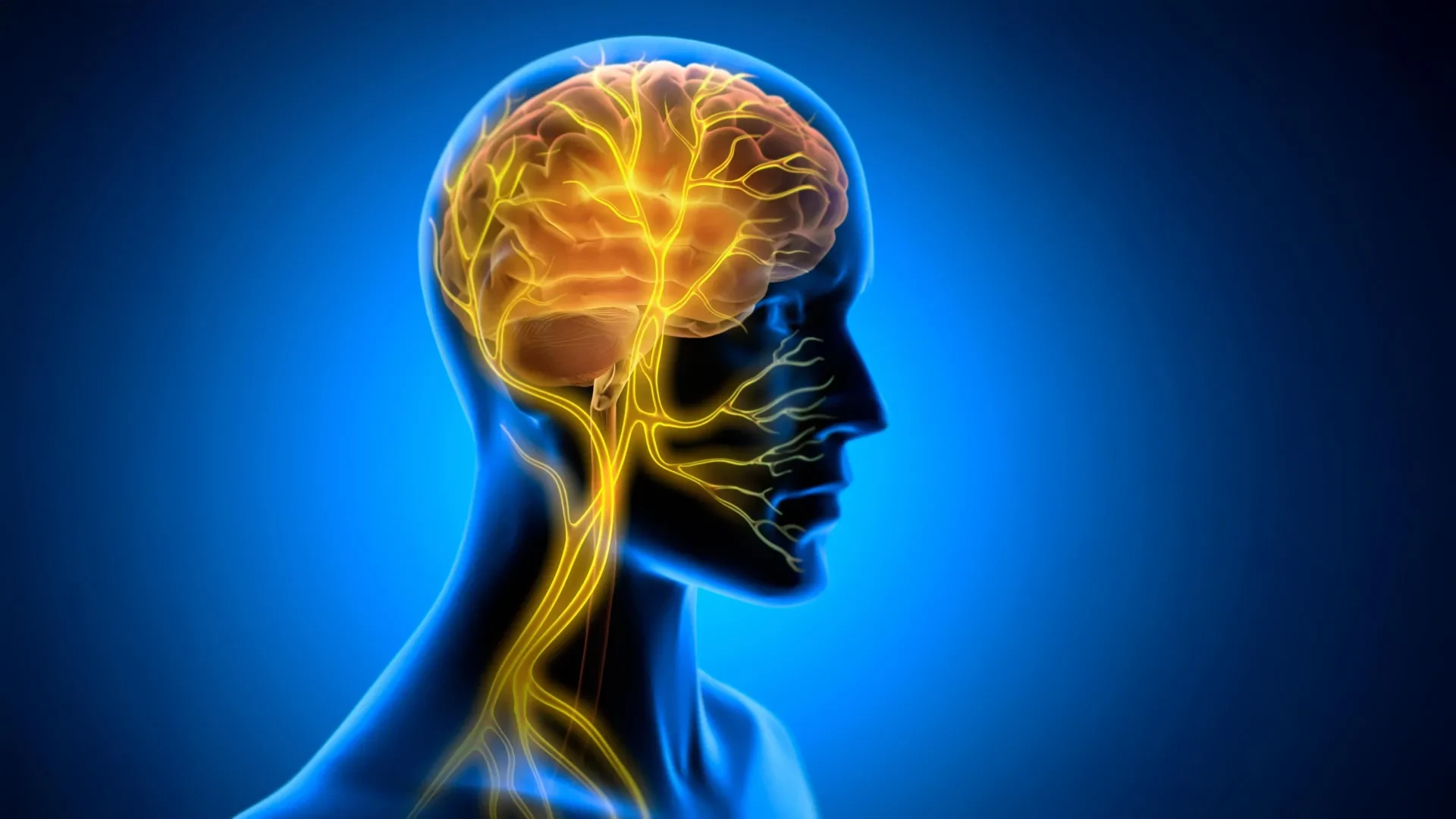
To understand the profound implications of the Tohoku University study, one must first examine the vagus nerve itself. Serving as one of the most critical communication pathways in the entire nervous system, the vagus nerve functions as a massive, bidirectional information highway linking the brain with nearly all of the body’s major internal organs, including the heart, lungs, and digestive tract. It continuously carries vital physiological data upward from the organs to the brainstem and higher brain centers, while simultaneously transmitting regulatory instructions downward from the brain to keep those internal systems functioning harmoniously.
Because of its extensive reach and powerful influence over central nervous system activity, scientists have long studied ways to influence this pathway. Vagus nerve stimulation, commonly known as VNS, involves delivering mild electrical impulses to the nerve. In modern medicine, VNS is already a clinically approved therapeutic approach utilized for treating several severe disorders, most notably drug-resistant epilepsy and treatment-resistant depression.
Historically, neuroscientists studying the cognitive effects of VNS have primarily focused on its capacity for neuromodulation. Under this conventional framework, electrical impulses delivered to the vagus nerve travel upward to trigger the release of vital neurotransmitters—such as norepinephrine, serotonin, and acetylcholine—across various regions of the brain. These chemical messengers are known to alter overall neural activity and heighten general arousal or attention. However, the new study from Tohoku University suggests that neurotransmitter release may only be part of a much larger, more sophisticated biological puzzle. The researchers suspected there might be another crucial mechanism at play: rhythmic, localized changes in the blood vessels residing deep inside the brain tissue itself.
To investigate this hypothesis and test whether bodily signals could directly influence motor skill acquisition, the research team designed a specialized piece of experimental hardware. They created a miniature cuff electrode specifically engineered to remain securely attached to the left cervical vagus nerve in living mice without causing discomfort or impairing natural movement. With this apparatus in place, the scientists subjected the test subjects to horizontal optokinetic response learning, widely known as HOKR learning.
HOKR is a classic, cerebellum-dependent eye movement task frequently used in neuroscience laboratories to evaluate motor learning and adaptation. During the task, mice are exposed to moving visual stripes, which trains them to instinctively improve their ability to track the visual field. This involuntary tracking response is functionally analogous to the automatic eye movements a human commuter makes while standing on a railway platform and watching a series of trains or scenery blur past. It relies heavily on precise neural coordination within the cerebellum, the region of the brain primarily responsible for motor control, balance, and fine-tuning physical actions.
The Biggest Effect Appeared After Practice
One of the most surprising and revealing aspects of the Tohoku University study involved the precise timing of the intervention. Rather than applying vagus nerve stimulation continuously during the physical training task itself, the researchers chose to deliver the stimulation strictly after each practice session had concluded.
When the mice were actively engaged in the HOKR learning task, applying VNS produced no immediate, observable boost in performance. The animals did not track the moving visual stripes any better during the stimulation than they did during standard, unassisted practice sessions. Instead, the true benefits of the nerve stimulation remained hidden during the practice phase and only became fully apparent much later.
When the researchers evaluated the animals on subsequent days, a striking divergence emerged. Mice that had received VNS immediately following their training sessions consistently demonstrated significantly stronger long-term learning outcomes compared to control groups that underwent training without the subsequent nerve stimulation. This distinct chronological pattern strongly implies that vagus nerve activation does not directly alter the raw execution of a motor task in the moment. Rather, it appears to profoundly influence the crucial post-training consolidation window—the delicate biological period during which the physical brain stabilizes, processes, and cements newly acquired information into durable, long-term memory structures.
"The key point is that VNS was delivered only after training," notes Professor Ko Matsui, a principal investigator on the research team. "Our findings suggest that VNS may open a hidden window of opportunity for enhanced learning by making the brain environment more receptive to long-lasting change."
This insight fundamentally shifts how scientists view the mechanics of practice. It suggests that the physiological state of the body in the immediate aftermath of learning is just as critical to skill retention as the focus and effort expended while the task is actually being performed.
Rhythmic Changes in Brain Blood Volume
Driven by the realization that VNS exerts its most powerful effects after training has ceased, the research team sought to uncover the specific physiological changes occurring inside the brain that might accompany this enhanced learning capacity. Specifically, they decided to measure local blood volume activity located in close proximity to the cerebellar flocculus, a specialized subregion of the cerebellum known to be directly involved in mediating HOKR learning and visual-motor coordination.
To capture these subtle internal shifts, the scientists utilized an advanced optical technique known as fiber photometry. This method allowed them to monitor real-time physiological dynamics within targeted neural tissues with remarkable precision.
The fiber photometry data revealed a fascinating biological chain of events. A single, discrete round of vagus nerve stimulation produced a distinct, two-phase vascular response within the cerebellar region. Immediately following the stimulation, local blood volume in the area experienced a brief, transient decrease. This initial reduction was systematically followed, after a slight delay, by a pronounced increase in blood volume.
Crucially, when the researchers administered VNS repeatedly—mimicking a structured protocol of post-training intervention—these localized vascular shifts did not remain isolated events. Instead, they began to pattern themselves into distinct, rhythmic oscillations in blood volume across the brain tissue.
Furthermore, these vascular rhythms were not merely an interesting biological byproduct; they appeared to be fundamentally tied to the success of the learning process itself. When the researchers analyzed the behavioral data, they discovered that individual mice which exhibited larger, more robust blood volume oscillations consistently achieved superior learning scores by the fifth day of the experiment. This strong correlation suggested a direct link between the dynamic, vascular environment of the brain and the enduring, long-term behavioral effects induced by vagus nerve stimulation.
"Our brains may be more strongly influenced by the body than we imagine," observes lead author Junyu Chen. "By tuning the brain’s metabolic environment, including rhythmic vascular movements, we may eventually unlock capacities that would otherwise remain latent."
Exploring the Brain-Body Connection
The revelation that rhythmic vascular changes in the brain can be successfully orchestrated by stimulating a peripheral nerve in the body opens up wide-ranging questions for future scientific investigation. While the current study provides compelling evidence from animal models, it also establishes a clear roadmap for the next phases of research in the field of neurophysiology.
Moving forward, the research team at Tohoku University plans to focus heavily on refining their stimulation protocols. This will involve testing different frequencies, intensities, and durations of vagus nerve stimulation to determine the exact parameters required to maximize post-training plasticity. Additionally, scientists aim to map out more precisely the intricate molecular and cellular pathways through which communication originating in the internal organs ultimately supports long-term structural changes within cerebellar networks.
By studying this complex, two-way communication loop between the brain and the body in greater detail, researchers hope to gain a much deeper comprehension of how transient experiences are permanently etched into memory. Ultimately, these insights could pave the way for novel therapeutic approaches and training enhancements designed to help humans acquire vital physical and cognitive skills more efficiently, ensuring that what is practiced today becomes a permanent capability for tomorrow.