
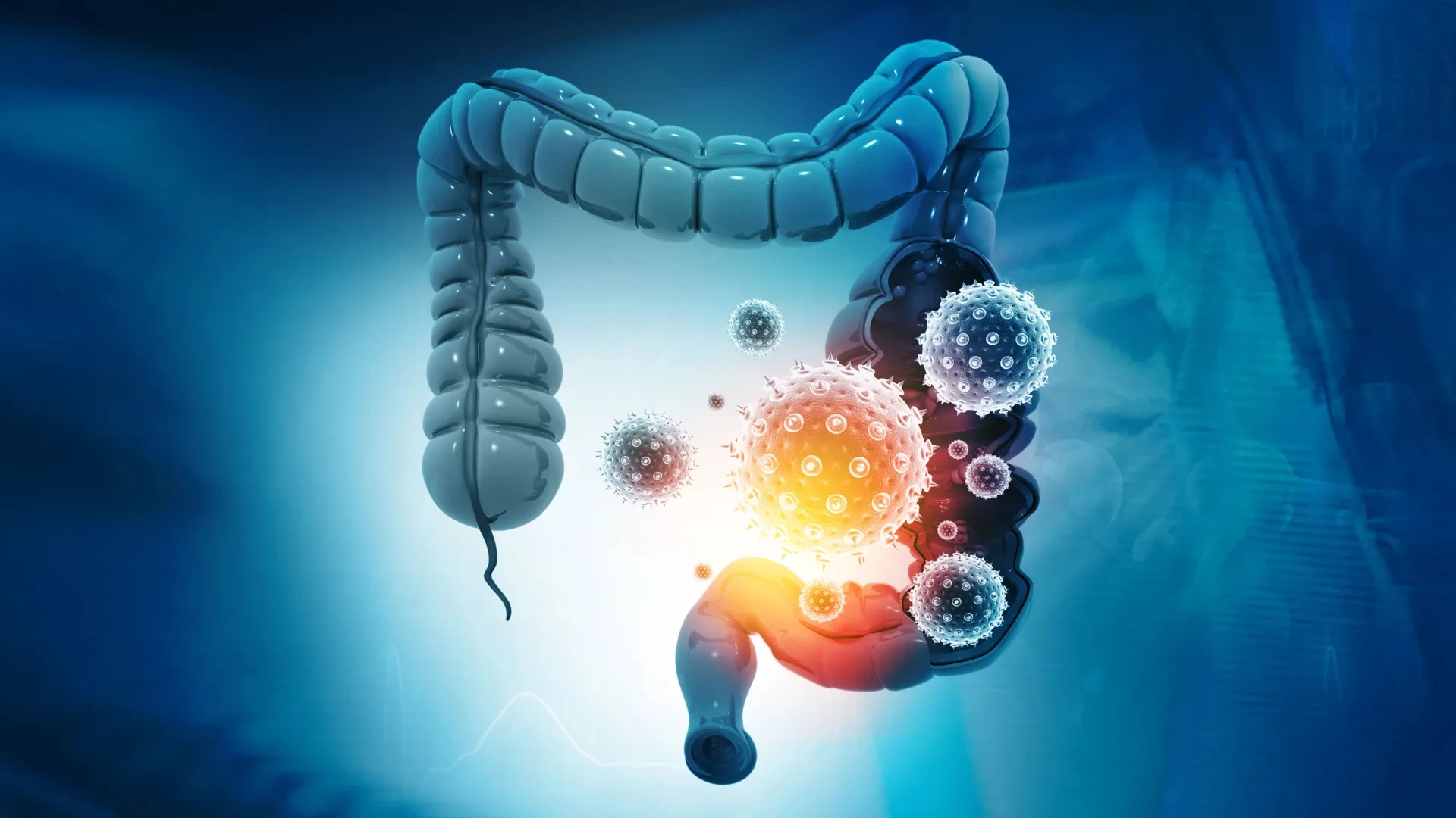
For nearly two decades, scientists have recognized that a common bacterium residing quietly within the human gut can produce a potent toxin capable of damaging the colon and driving the formation of tumors. Yet, despite extensive research into the mechanics of this process, one critical piece of the puzzle remained persistently out of reach: no one could explain how the toxin first attaches to intestinal cells to initiate its destructive assault.
Now, an international team of researchers has successfully uncovered that missing step. By identifying the precise cellular gateway utilized by the bacteria, the scientists have revealed a surprising biological vulnerability that could ultimately pave the way for entirely new methods of protecting the colon, preventing dangerous infections, and fighting colorectal cancer.
The breakthrough is the result of a collaborative effort led by investigators at the Johns Hopkins Kimmel Cancer Center, the Bloomberg~Kimmel Institute for Cancer Immunotherapy, and the Johns Hopkins University School of Medicine. Their findings, published in the prestigious scientific journal Nature, pinpoint a specific protein known as claudin-4 as the essential docking station for a bacterial toxin called BFT.
Even more promising than the identification of the receptor itself is how quickly the research team translated the discovery into a tangible defensive strategy. The scientists have already successfully designed a molecular decoy engineered to intercept the toxin before it can reach or damage vulnerable colon cells. In laboratory tests involving mice, this protective strategy successfully shielded the animals from the toxin’s harmful biological effects, pointing toward a possible future approach for preventing the bacterial tissue damage long associated with colorectal cancer. The research received partial financial support from the National Institutes of Health, alongside contributions from several other major scientific foundations and institutions.
"We’ve made several attempts over time to identify the receptor, so this is an exciting moment," says senior author Cynthia Sears, M.D., the Bloomberg~Kimmel Professor of Cancer Immunotherapy and a professor of medicine at Johns Hopkins. "Understanding how bacterial toxins work can open doors to new approaches for detection and therapy for associated diseases, including diarrhea, colorectal cancer and bloodstream infections."
How a Common Gut Bacterium Can Promote Colon Cancer
The investigation centers on Bacteroides fragilis, a remarkably common microorganism that can be found thriving naturally in the intestines of up to 20 percent of healthy people. Under normal circumstances, the presence of this bacterium does not automatically mean an individual will develop cancer or experience chronic illness. However, specific strains of the organism produce a powerful toxin capable of disrupting the delicate protective lining of the colon, triggering localized inflammation, and actively encouraging abnormal tumor growth over time.
This specific toxin, formally known as B. fragilis toxin or BFT, has been under intense scientific scrutiny for many years. In a landmark 2009 study published in Nature Medicine, Dr. Sears and her colleagues first demonstrated that the toxin possessed the alarming ability to promote the development of colon tumors in experimental settings. That foundational work helped establish a vital clinical and biological connection between toxin-producing gut bacteria, chronic intestinal inflammation, and the progression of colorectal cancer.
Earlier biochemical experiments also revealed that BFT systematically damages a critical cellular protein called E-cadherin. Under normal conditions, E-cadherin acts like a molecular glue, helping to hold neighboring epithelial cells tightly together and maintaining the sturdy, impermeable barrier that protects the colon wall from harmful luminal contents. When this essential protein is fractured and broken apart by the advancing toxin, the protective intestinal barrier becomes severely compromised. This breakdown potentially allows various other harmful substances, toxins, and microbes to leak through and interfere with the sensitive surrounding tissue, accelerating disease processes.
However, even as researchers documented the destruction of E-cadherin, a major paradox remained completely unresolved. Although BFT was clearly responsible for breaking down E-cadherin, laboratory observations showed that the toxin did not appear to attach directly to that specific protein. Investigators long suspected that an entirely separate molecule must be acting as an intermediary, helping the toxin latch onto the cell surface and reach its target, but repeated attempts by various laboratories to identify this elusive molecule had consistently failed.
Scientists Identify the Missing Link Using CRISPR
To solve a biological mystery that had persisted for nearly twenty years, the research team decided to leverage CRISPR, a cutting-edge gene-editing technology that allows scientists to systematically disable individual genes within living cells to investigate their precise functions and determine their roles in complex biological pathways.
Maxwell White, an M.D./Ph.D. candidate working directly within Dr. Sears’s laboratory, spearheaded a comprehensive genome-wide CRISPR screening effort in close collaboration with the laboratory of Matthew Waldor at Harvard Medical School. The research team systematically switched off thousands of individual genes within colon epithelial cells—the specialized cells that form the continuous inner lining of the colon—to determine precisely which genetic components were strictly necessary for the bacterial toxin to successfully attach and inflict cellular damage.
As the massive screening data was analyzed, one specific gene stood out dramatically from all the rest. The gene is responsible for producing claudin-4, a well-known protein that helps neighboring epithelial cells form tight junctions. These tight junctions act as microscopic seals that strictly limit the uncontrolled movement of fluids and substances between cells, thereby playing a fundamental role in preserving the structural integrity of the intestinal barrier.
When the researchers experimentally disabled the specific gene responsible for producing claudin-4, the results were definitive: BFT could no longer bind to the surface of the colon cells. Deprived of that initial physical attachment point, the toxin was rendered entirely incapable of reaching or damaging E-cadherin, effectively halting the pathogenic cascade before it could begin.
The finding definitively proved that claudin-4 functions as the long-sought primary receptor for the bacterial toxin. In effect, the toxin must first latch firmly onto this specific protein framework on the cell membrane before it can carry out any of its subsequent damaging biological activities.
"It took a while to get the assay working and validate the approach, but once we were able to do the screen, claudin-4 was a clear, resounding top hit," says White. "That was an exciting moment."
An Unexpected Discovery Reveals a New Bacterial Attack Strategy
The exact identity of the receptor came as a significant surprise to the scientific team. Prior to the genome-wide screen, Dr. Sears and other leading researchers in the field had fully expected BFT to interact with a protein actively involved in complex cellular signaling pathways, such as a G protein-coupled receptor. These types of cell-surface receptors typically act like molecular communication switches, allowing cells to sense and respond to chemical signals from their surrounding environment.
Claudin-4, however, serves a profoundly different structural purpose within the body. Rather than functioning primarily as a signaling receptor, its main physiological job is to help maintain the robust physical connections and tight seals between neighboring cells in epithelial tissues.
Furthermore, the discovery exposed an unusual and previously unappreciated mechanism of action for a bacterial toxin. BFT belongs to a broader class of biological enzymes known as proteases, which are specialized proteins designed to cut and degrade other proteins. While the vast majority of known proteases act directly and immediately upon their primary targets, BFT appears to require an entirely separate structural receptor to anchor itself to the cell membrane before it can reach, access, and successfully damage E-cadherin.
When the research team reviewed the existing body of scientific literature, they could not identify any other bacterial toxin known to utilize this exact two-step binding and cleavage mechanism.
To definitively establish that BFT and claudin-4 genuinely bind to one another in physical reality, the Johns Hopkins researchers partnered with structural biologists F. Xavier Gomis-Rüth and Ulrich Eckhard at the Molecular Biology Institute of Barcelona. Utilizing advanced biophysical techniques designed to measure precise molecular interactions, White and the Barcelona team demonstrated conclusively that BFT and claudin-4 form a tightly bound biochemical complex in laboratory experiments. Each individual molecule of the toxin paired directly with one molecule of the receptor, providing concrete physical evidence of the connection.
A Clever Molecular Decoy Blocks the Dangerous Gut Toxin
Identifying the specific cellular receptor did much more than simply explain the mechanical details of how the toxin operates; it also immediately provided the scientists with a brilliant conceptual framework for stopping it.
Reasoning that if BFT strictly requires claudin-4 to attach to colon cells in the first place, the researchers hypothesized that a carefully engineered substitute could be introduced to safely lure the toxin away from its intended target within the human body.
To test this ambitious hypothesis, the team engineered a specialized molecular decoy. This decoy consisted of a soluble protein carrying precise structural sequences derived from claudin-4, purposefully designed to attract and trap BFT without allowing the toxin to launch an attack on the colon’s protective cellular lining.
The researchers then teamed up with the laboratory of Min Dong at Harvard Medical School, including investigator Kang Wang and fellow colleagues, to test the experimental approach in living animal models. These in vivo experiments allowed the scientists to observe how the bacterial toxin behaves within the vastly more complicated physiological environment of a living mammalian intestine.
The experimental results proved remarkably encouraging. Instead of binding to the natural claudin-4 receptors located on the surface of the colon cells, the circulating BFT preferentially attached itself to the introduced decoy proteins. By successfully intercepting and neutralizing the toxin before it could make contact with intestinal tissue, the molecular decoys reliably protected the mice from experiencing BFT-induced cellular damage.
The finding strongly suggests that future therapeutic interventions might successfully focus on interfering with the toxin’s very first step—its initial attachment phase—rather than attempting to repair the extensive tissue damage after it has already occurred.
"This approach could be iterated upon with small molecules or other biologics that have better pharmacological properties," says White. The collaborative research team is already actively exploring which specific molecular approaches might prove most successful in developing clinical-grade blockers for the toxin.
Small molecules represent chemical compounds that can frequently be developed into stable, orally administered medicines, while biologics are advanced treatments manufactured from biological materials, such as engineered proteins. Future iterations of this decoy strategy could potentially be optimized to circulate more effectively within the human body or be administered with greater clinical ease.
What This Discovery Could Mean for Colon Cancer Prevention
Although these new findings successfully identify a promising and actionable therapeutic target, important scientific questions naturally remain to be answered through future research.
While the investigators have definitively established that BFT attaches to claudin-4 and that blocking this specific molecular interaction can protect living mice from toxin-induced tissue damage, they have not yet managed to capture the exact experimental three-dimensional structure showing precisely how the two molecules fit together at an atomic level.
Notably, even state-of-the-art artificial intelligence tools, including advanced protein-modeling software like AlphaFold, have thus far been unable to fully resolve the precise structural nature of this interaction. Achieving a complete understanding of this 3D structure could ultimately help researchers design even more potent and specific compounds capable of preventing the toxin from binding.
Further preclinical and clinical experiments will also be strictly required to determine whether this innovative decoy approach can safely and effectively prevent toxin-related diseases in human patients. While the current mouse experiments successfully demonstrated protection against acute tissue damage, they did not establish whether the strategy can outright prevent the long-term development of colorectal cancer.
Nevertheless, the discovery successfully resolves a major scientific question that has persisted ever since the original connection between BFT and colon tumor formation was first established back in 2009. By finally identifying the toxin’s previously elusive point of attachment, the researchers have uncovered a viable, highly promising way to interrupt a dangerous biological disease process right at its inception.
Ultimately, these new insights could support the development of entirely novel diagnostic and therapeutic strategies aimed at detecting, preventing, or treating a variety of severe conditions associated with bacterial toxins, including colorectal cancer, persistent diarrhea, and dangerous bloodstream infections.
Additional researchers who contributed significantly to the published study include Jason Chen, Shaoguang Wu, Abby L. Geis, and Jessica Queen of Johns Hopkins, alongside Hailong Zhang, Karthik Hullahalli, and Jie Zhang of Harvard Medical School.
Financial support for the research was provided by the Bloomberg~Kimmel Institute for Cancer Immunotherapy, Janssen Research and Development, Cancer Research UK, the National Institutes of Health under specific grant numbers R01 AI042347, R01 NS080833, R01 NS117626, R01 AI170835, and R01 AI189789, as well as the Howard Hughes Medical Institute.